
Insulin synthesis and secretion are the linked processes that let pancreatic beta cells make insulin, package it, and release it when blood glucose rises. Beta cells first build the hormone from precursor proteins, convert proinsulin into insulin and C-peptide, store both in secretory granules, and then use glucose-sensitive ion channels and calcium signals to trigger release. This matters because a problem at any step can disrupt glucose control and contribute to diabetes.
At a high level, the pathway starts in the pancreas, inside the islets that contain beta cells. From there, glucose sensing, cellular metabolism, electrical signaling, and granule exocytosis work together as one coordinated system. The sections below explain where insulin is produced, how blood glucose triggers release, what first-phase and second-phase secretion mean, and where this biology fits into everyday diabetes care.
Key Takeaways
- Beta cells in pancreatic islets make insulin as preproinsulin, then proinsulin.
- Proinsulin is processed into mature insulin and C-peptide before storage.
- Glucose metabolism raises ATP, closes KATP channels, and opens calcium channels.
- Calcium entry triggers granule exocytosis, which releases stored insulin.
- Beta-cell dysfunction can impair insulin production, insulin release, or both.
CanadianInsulin.com operates as a prescription referral platform.
How Insulin Synthesis and Secretion Begin
Insulin is made in pancreatic beta cells, which sit inside the islets of Langerhans. These small endocrine cell clusters are central to the physiology behind the Diabetes Hub. The process starts when the insulin gene is transcribed and translated into preproinsulin, an early precursor that contains a signal sequence directing it into the endoplasmic reticulum.
Inside the cell, that signal sequence is removed, leaving proinsulin. Proinsulin folds into its correct shape and forms disulfide bonds, which are critical for a functional insulin molecule. It then moves through the Golgi apparatus into immature secretory granules, where enzymes cleave proinsulin into mature insulin and C-peptide. Both are packaged together and stored until the cell receives the right signal to release them.
| Stage | What Happens | Why It Matters |
|---|---|---|
| Preproinsulin | The beta cell first translates the insulin precursor with a signal peptide. | This directs the new protein into the cell machinery for hormone processing. |
| Proinsulin | The signal peptide is removed and the protein folds into a stable intermediate. | Proper folding is needed before insulin can be activated and stored. |
| Mature insulin | Granule enzymes cut proinsulin into insulin and C-peptide. | The hormone is now ready for storage and regulated release. |
This is not a one-time event. Beta cells continually adjust insulin gene expression, protein synthesis, granule formation, and storage based on metabolic demand. In healthy physiology, the same cell must make enough hormone to meet future need while keeping a pool ready for rapid release after meals.
The Glucose-Triggered Release Pathway
When blood glucose rises, the beta cell converts a nutrient signal into an electrical signal and then into hormone release. This sequence is often called stimulus-secretion coupling, which means the cell can sense a change in fuel availability and translate it into exocytosis, the fusion of insulin granules with the cell membrane.
Glucose enters the beta cell through glucose transporters and is rapidly metabolized. As glucose metabolism increases, the ATP-to-ADP ratio rises. That closes ATP-sensitive potassium channels, often shortened to KATP channels. The cell membrane then depolarizes, voltage-dependent calcium channels open, and calcium flows into the cell. That calcium surge is the immediate trigger for granule fusion and insulin release.
- Glucose entry – glucose moves into the beta cell.
- Metabolic sensing – glucose breakdown raises cellular ATP.
- KATP closure – potassium efflux falls as the channel shuts.
- Membrane depolarization – the beta-cell membrane becomes electrically activated.
- Calcium influx – voltage-gated calcium channels open.
- Granule exocytosis – stored insulin is released into the bloodstream.
This pathway explains why insulin secretion is closely tied to nutrient metabolism, not just the presence of glucose in the blood. The speed of carbohydrate absorption, the mix of nutrients in a meal, and hormonal signals from the gut can all shape how strongly the pathway is activated. That is one reason meal-response topics such as the Food Insulin Index often come up in diabetes education.
First-Phase and Second-Phase Secretion
Insulin release usually occurs in two broad phases. First-phase secretion is the rapid burst that comes from granules already docked and ready at the membrane. Second-phase secretion is the slower, sustained release that depends on recruitment of reserve granules, continued metabolism, and ongoing signaling inside the cell.
That distinction matters because early type 2 diabetes often weakens first-phase secretion before total insulin production is lost. As a result, the body may struggle with post-meal glucose spikes even when some insulin output is still present.
Why it matters: Timing is as important as quantity in normal insulin release.
What Beta-Cell Control Really Means
Beta-cell control is broader than one ion channel or one hormone. It includes insulin gene expression, protein folding, proinsulin processing, mitochondrial metabolism, membrane excitability, granule trafficking, and exocytosis. If any part of that chain becomes inefficient, insulin output may fall or become poorly timed.
Hormonal signals add another layer of control. Incretin hormones, especially GLP-1 and GIP, can amplify glucose-dependent insulin secretion after meals. For broader context, see GLP-1 Explained and GLP-1 Basics. Neural input matters too. Parasympathetic signaling can support release around meals, while somatostatin from nearby delta cells generally restrains secretion.
Control also happens over longer time scales. When tissues become insulin resistant, beta cells often try to compensate by making and releasing more insulin. At first, that adaptation can preserve glucose control. Over time, however, persistent metabolic stress may impair mitochondrial function, disrupt granule handling, and reduce the cell’s ability to maintain normal insulin synthesis and secretion.
Why C-Peptide and Secretion Phases Matter
C-peptide is the connecting segment removed when proinsulin is converted into mature insulin. Because beta cells release endogenous insulin and C-peptide together, the presence of C-peptide can provide a rough sense of how much insulin the pancreas is still making. Injected insulin does not include C-peptide, so the two are not interchangeable markers.
Storage dynamics matter as much as processing. Beta cells maintain a readily releasable pool of granules near the membrane and a larger reserve pool deeper in the cell. Proteins that control docking, priming, and membrane fusion help decide how quickly insulin can be released once calcium enters. Calcium is the final trigger, but it does not work alone. ATP generation, cAMP signaling, and cytoskeletal transport all shape how much hormone actually reaches the cell surface.
- Islet – a hormone-producing cell cluster in the pancreas.
- Stimulus-secretion coupling – conversion of nutrient sensing into insulin release.
- KATP channel – an ATP-sensitive potassium channel that helps set membrane voltage.
- Exocytosis – fusion of an insulin granule with the cell membrane.
Put simply, insulin biosynthesis answers how the hormone is made, while secretion physiology answers how it is released at the right moment. The body needs both systems to work together. Strong production with poor release timing is not enough. Fast signaling with depleted hormone stores is not enough either.
What Goes Wrong in Diabetes
In diabetes, beta cells may be absent, injured, overworked, or less responsive to glucose. In type 1 diabetes, autoimmune destruction sharply reduces or eliminates endogenous insulin production. In type 2 diabetes, insulin resistance raises demand, and beta cells may initially compensate by increasing output before losing precision and capacity over time.
Early dysfunction often shows up as weaker first-phase secretion, delayed meal responses, or incomplete proinsulin processing. Chronic high glucose, excess fatty acid exposure, inflammation, oxidative stress, and inherited susceptibility can all contribute. That helps explain why some people first notice post-meal spikes or fatigue before they understand the underlying beta-cell problem. For related warning signs, see High Blood Sugar Symptoms.
Abnormal glucose patterns do not all reflect the same mechanism. Some episodes relate to impaired endogenous secretion, some to insulin resistance, and some to treatment effects or meal timing. A separate review of Reactive Hypoglycemia explains why low-sugar symptoms after meals can have several causes. For broader background, the Diabetes Articles collection brings together condition, symptom, and treatment topics.
How This Science Fits Into Diabetes Care
This physiology matters because diabetes treatments do not all act on the same step. Some therapies mainly improve insulin sensitivity or reduce glucose production by the liver. Others act more directly on the beta cell, either by enhancing glucose-dependent secretion or by stimulating release more directly.
For example, metformin is generally used to improve glucose handling without forcing a strong pancreatic burst. By contrast, meglitinides can stimulate insulin release from beta cells, which is why mechanisms matter when comparing treatment classes. A neutral overview of that pathway is available in Repaglinide Uses. GLP-1-based treatments work through incretin signaling and are best understood in the context of glucose-dependent insulin secretion rather than simple replacement.
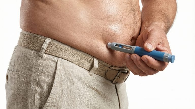
When endogenous insulin production becomes too low, external insulin replaces what beta cells can no longer supply. At that point, the practical questions shift from synthesis to delivery and technique. Related reading includes Insulin Pump Basics, Injection Sites, and Lipohypertrophy. If you are comparing device or medication categories, the browseable Diabetes Products hub gives category-level context.
Quick tip: When reading about a diabetes drug, ask whether it improves sensitivity, boosts secretion, or replaces insulin.
Licensed third-party pharmacies handle dispensing where permitted.
Authoritative Sources
- For a deeper review of beta-cell physiology, see the NIH-hosted review on regulation of insulin synthesis and secretion.
- For stepwise background on hormone formation, use the NCBI Bookshelf chapter on insulin biosynthesis and secretion.
- For meal-related signaling pathways, review the Pancreapedia article on insulin release in response to diet and hormones.
Insulin synthesis and secretion depend on healthy beta cells, accurate glucose sensing, intact granule processing, and calcium-triggered exocytosis. Once that chain weakens, glucose control can change quickly. Further reading can help connect the cellular pathway to symptoms, treatment classes, and day-to-day diabetes management.
This content is for informational purposes only and is not a substitute for professional medical advice.